Detectors
SECONDARY ELECTRON DETECTORS, IMAGE QUALITY & CONTRAST
Lecture and presentation for Microscopy Society of America August 1998
David Scharf Scharf Microscopy, Los Angeles, CA 90039 The Secondary Electron Detector (SED) (Figure 1.) is the first link in the chain of image formation after the initial illumination of a specimen in a Scanning Electron Microscope (SEM), therefore it is most crucial that the efficiency of the SED be as great as possible in order to maximize Signal-to-Noise Ratio (S/N) as any imperfections, inadequacies, and noise are then amplified along with the signal by the subsequent electronic amplifiers in the signal chain. In the Everhart-Thornley SED, matching the scintillator’s output spectrum to the photomultiplier tube’s photocathode spectral sensitivity is one factor along with other characteristics of the PM tube, like quantum efficiency and noise characteristics contributing to performance. Lightpipe material along with as optical coupling techniques (Figure 2.) are another factor which determines the overall efficiency of converting SE electron strikes on the scintillator to a video output signal. We typically use P47 phosphor scintillators (Figure 3.) coupled to 10mm quartz lightpipes feeding to a 52mm PM tube having bi-alkalai photocathode (Figures 4. & 5.) with reduced area (10mm), such as the Thorn-EMI 9844B. The position of the SED in relation to the sample is important in determining collection efficiency and contrast. At higher magnifications, the ratio of sample dimensions to the distance to the detector is very high, but at low magnifications the sample dimensions can become a significant portion of the sample-to-SED distance, thereby creating greater overall sample contrast. This particular kind of macro-contrast can be detrimental to overall image quality because the microcontrast of small features cannot be maximized without saturating the highlights of portions of the image. This kind of imaging problem can be minimized by moving the detector position further away from the sample. But this also maximizes collection of “stray” signals from distant chamber structures. At medium and high magnifications, we keep our detector position very close to the sample to maximize collection efficiency. Simultaneous use of multiple detectors is another way to manipulate image contrast. Traditionally, the SED is placed in a position so that the sample is tilted towards it, thus aiding in signal collection. Placing additional detectors in other positions can supply other important image information which may then be used to create Color Contrast [1]. Two SEM setups were used to investigate this work on detectors. An ETEC Autoscan with 3 variable bias SEDs at 30° horizontal separation and a LEO 440 with 3 SEDs similarly configured. For optimum S/N, SE emission from the sample is proportioned to each SED by varying the individual bias of each detector. Three different images are formed simultaneously, whereupon the individual video signals are encoded with a color (by dividing into R.G.B. components) then combining them into one color image, thus allowing real-time viewing of a Color-Contrast Encoded image. Contrast is produced from having a different color coming from the position of each respective SED. This technique supplies significant additional information regarding surface topography and is particularly effective for reading ambiguous surface features. Proper utilization of multiple detectors can result in a slight improvement in collection efficiency. This technique should also work where one detector is a through-the-lens SED.
References 1. D. Scharf, U.S. Patent No. 5,212,383 (1993)
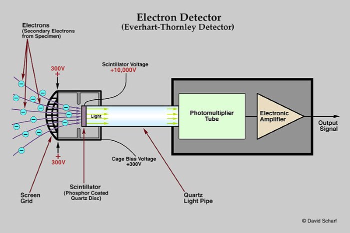
Figure 1. Everhart-Thornley seconday electron detector
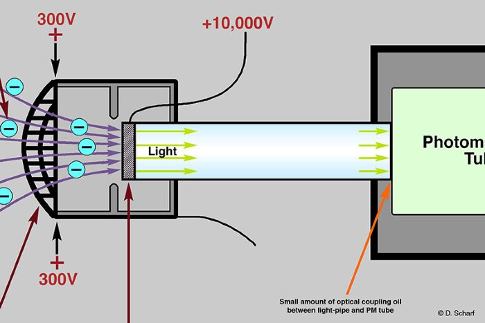
Figure 2. Secondary electron detector detail
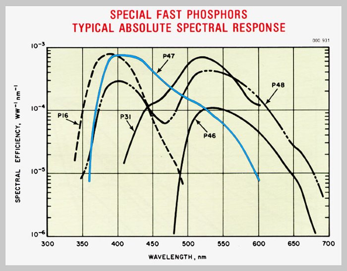
Figure 3. Phosphors for scintillator
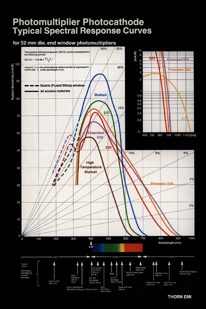
Figure 4. PMT Photocathode response curves
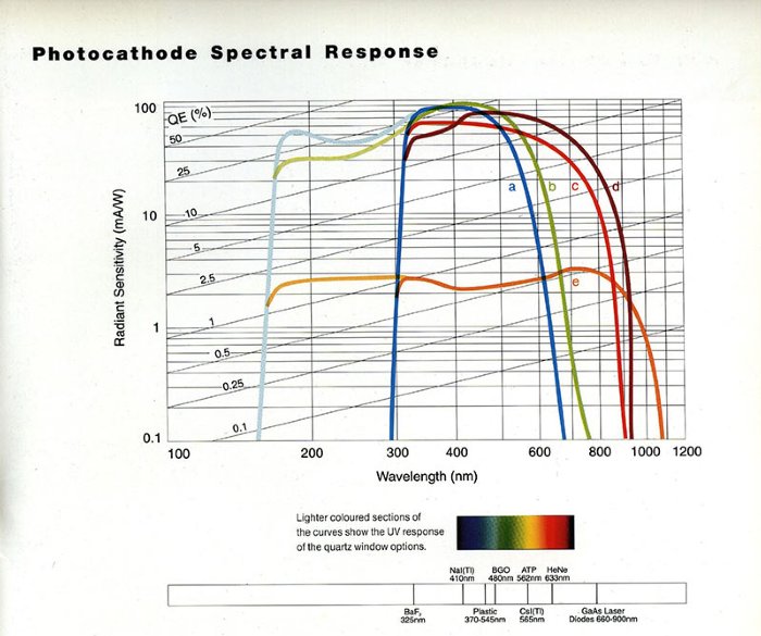
Figure 5. Photocathode spectral response; Curve “a” is Bialkalai